The Imaging Centre

Core facility for advanced microscopy

Training and technical support in a variety of microscopy techniques, including light, confocal, scanning electron (SEM) and high-resolution scanning transmission electron microscopy (TEM). 

The Core Facility offers a comprehensive service, including imaging and custom sample preparation.

We can provide guidance and technical expertise on the use of our entire infrastructure, to provide a low threshold for all users whether experienced or not.

The Imaging Centre is accessible to all internal and external users, both academic and industrial.


Crossection of an Ash leaf

Contact information:

E-mail: imaging.centre@nmbu.no

Phone: 67 23 27 61/62, 92 41 72 43 or 48 25 98 43

Visiting address:
Oluf Thesens vei 43, 1433 Ås

See location in MazeMap.

Opening hours:

Kl. 08:00 to 15.45

15. May to 14. September:
kl. 08:00 to 15.00


  • News

    Flytting av Imagingsenteret utsettes /Relocation of Imaging Centre slightly postponed (sharepoint.com)

    Microscopy seminar on September 24th focusing on scanning electron microscopy and X-ray microscopy. The seminar is free and open to everyone. See more information in the link below.
    Welcome!

    ZEISS Seminar - SEM

  • Booking system and prices

    Prices: We have different prices for use of the various instruments; please contact us by e-mail, and we will arrange for a meeting or send you a price list. The University is using the Leiestedsmodellen, and the prices will be adjusted annually.

    From 01.01.24 there will be revised prices. For project applications that are submitted, it is important to use the correct prices. 

    Booking: The University is currently in the process of implementing a new booking system, but until this is in place please send us an e-mail or give us a call.


  • Courses

    BIO 300 Microscopy Techniques

    The course gives a thorough introduction to the use of light, confocal laser scanning, scanning- and transmission electron microscopy.

    We have lectures, demonstrations, exercises on general and special optics, sample preparation, staining methods and various imaging techniques.

    This course take place in the January block, and there will be given an oral exam in February/March.

    Sign up by sending an email to: imaging.centre@nmbu.no

    Read more about BIO 300.

    Please feel free to contact us if you have any questions.

    Light and confocal laser scanning microscopy

    Courses on demand

    We can arrange for courses specifically tailed for your needs in the different optical techniques. Please get in touch with us, and we can put together a course suited for your requirements.


  • Equipment

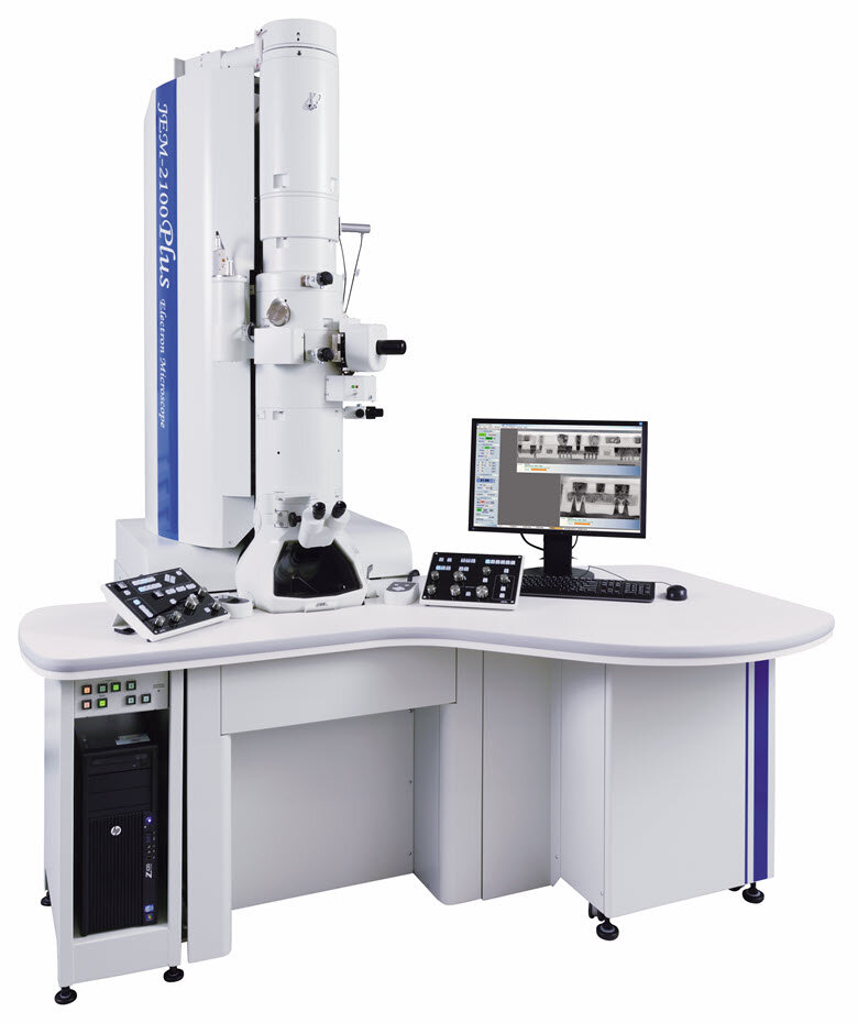
    Jeol Transmission electron microscope JEM 2100-Plus, TEM

    Jeol transmission electron microscope JEM-2100 plus with EDX

    The JEM-2100Plus is a multi purpose scanning transmission electron microscope, which combines the proven JEM-2100 optic system with an advanced control system for enhanced ease of operation.

    The microscope is equiped with a LaB6-filament, TVIPS camera, an EDS system from Oxford, BEI, DF and BF detectors. High voltage range 80 - 200kV.

    More about Jeol JEM 2100-Plus

    Zeiss EVO50 EP Scanning electron microscope, SEM

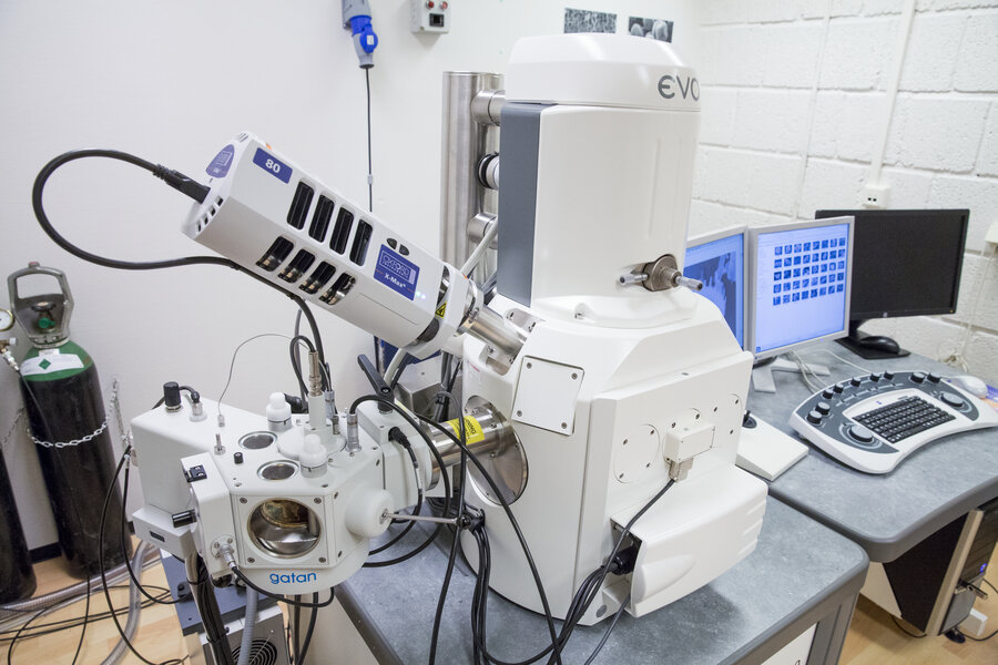
    Zeiss EVO 50 Scanning electron microscope

    Zeiss EVO 50 EP scanning electron microscope provides a flexible and productive tool for surface imaging at both high and low resolution.

    Our SEM is a Extended Pressure (EP) instrument where the specimen environment can be controlled and we can examine fresh samples.

    Detectors for Backscattered Electron Imaging (BEI) and EDS system makes it possible to detect and localize differences in element composition.

    More about the Zeiss EVO family.

    EDS system from Oxford

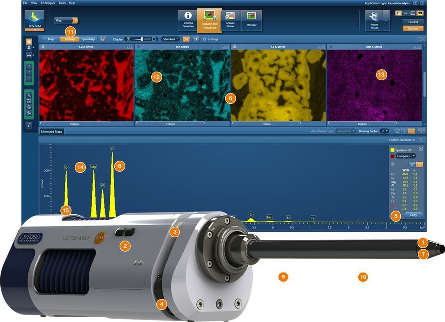
    EDS

    The EDS Analysis system provides elemental and chemical analysis of a sample inside the SEM and TEM.

    More about the EDS system.

    Zeiss Axio Zoom. V16 Stereomicroscope with fluorescent brightness

    Zeiss AxioZoomV16

    Axio Zoom.V16 combines a 16x zoom with a high numerical aperture of NA 0.25. It achieves a very high aperture in the medium zoom range, and you get superior brightness in large object fields.

    With Axio Zoom.V16, the fluorescence zoom microscope for large samples you can view complete model organisms in fluorescence contrast.

    The microscope is equiped with DAPI and GFP filtercubes.

    More about AxioZoomV16.

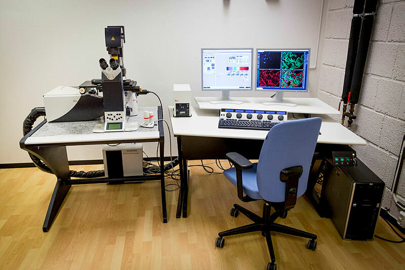
    Leica TCS SP 5 Confocal laser scanning microscope

    Leica SP5

    The Leica TCS SP5 Confocal covers a broad range of requirements in confocal and multiphoton imaging. The system provides the full range (spot recording to real-time imaging) of scan speed at high resolution.

    The TCS SP5 enables you to easily switch between conventional and resonant scanner.

    With high-efficiency SP detection (five channels simultaneously) and optional AOBS (Acousto Optical Beam Splitter), the system delivers bright, noise-free images with minimal photo damage at high speed.

    More about the Leica SP5.

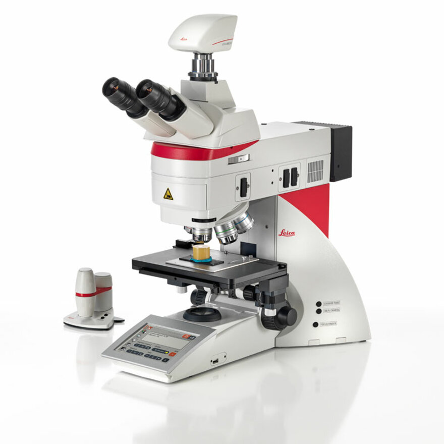
    Leica DM6B Light microscope

    Leica DM6B

    The Leica DM6B is a semi-automated research light microscope that provides all transmitted light contrast methods including automated differential interference contrast, polarization, dark field, phase contrast and fluorescense.

    Filter CubeExcitation (nm)Dichroic Mirror (nm)Emission (nm)
    DAP325-375400435-485
    UVG395-415495500-550
    GFP450-490495500-550
    N21515-561580LP 590

    The microscope is equipped with a DMC4500 Digital Camera, and the new LAS X analysis program.

    More about the Leica DM6B

    Leica LMD600 Laser micro dissection microscope

    Leica LMD600

    Leica LMD600 Laser Micro dissection microscope is a high-precision technology, for sample preparation to obtain well-defined starting material for downstream experiments in the fields of genomics, micro arrays, biochips, and proteomics.

    More about Leica laser micro dissection systems.

    Leica EM AFS2 and FSP Freeze substitution system

    Leica AFS 2

    Leica EM AFS 2 is an automatic freeze substitution system for embedding of samples following high pressure freezing and other cryo fixation methods, or progressive lowering of temperature (PLT) to substitute and resin infiltrate chemically fixed specimens.

    The sample is polymerized under UV light and can be cut and immunolabeled for transmission electron microscopy. It is equipped with the Leica EM FSP automatic reagent handling system.

    The Freeze substitution unit has an automatic reagent handling system, giving a one step preparation, reduced workload, and increased reproducibility.

    It minimizes operators contact with toxic media and reduces the risk of losing specimens.

    More about Leica EM AFS2.

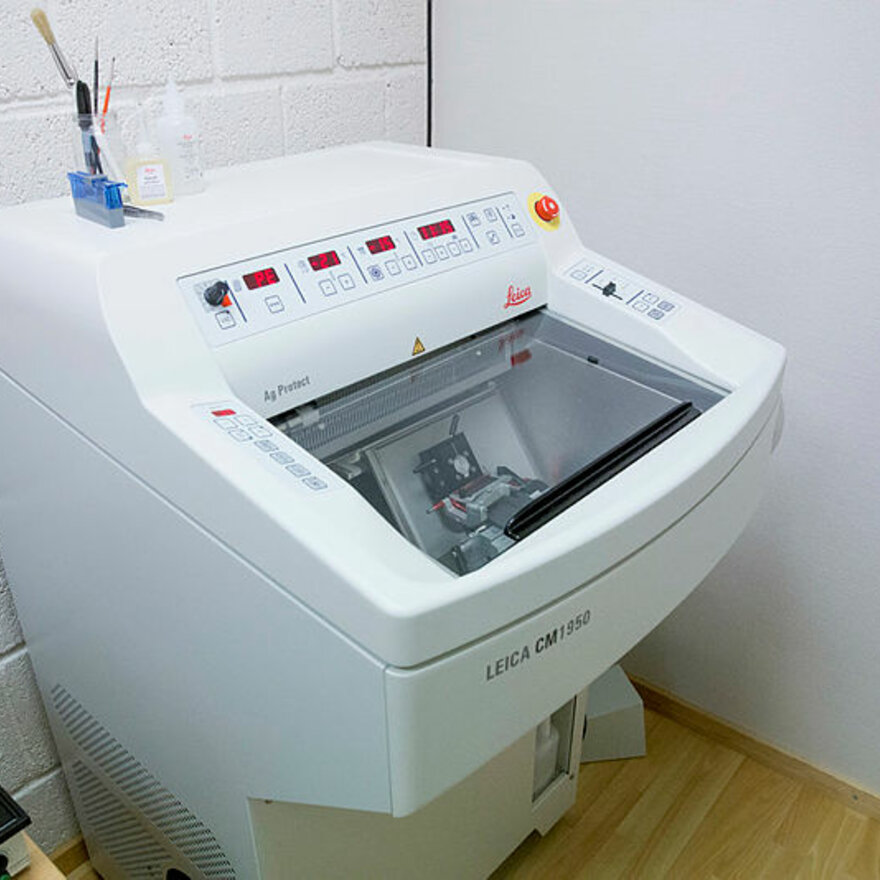
    Leica CM1950 Cryostat

    Leica Cryostat CM1950

    The Leica CM 1950 is a high quality motorized cryostat for sectioning at low temperatures. The sectionthickness and temperature can be precisely regulated, and adjusted to fit all kinds of samples.

    More about The Leica CM 1950

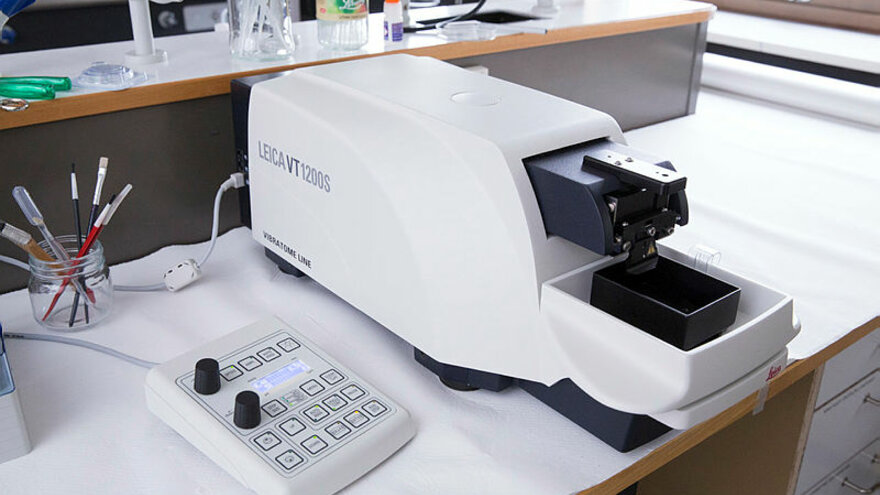
    Leica VT1200 S Vibratome

    Leica Vibratome

    The Leica VT1200 S is an automated vibratome. It has unique control and adjustments of unwanted vertical movements. You can section both fresh and fixed tissue.

    More about Leica VT1200 S Vibratome

    BAL-TEC CPD 030 Critical point dryer

    Kritisk punkt tørker

    The procedure of critical point drying is an efficient method for drying delicate samples for SEM applications. It preserves the surface structure of a specimen which could otherwise be damaged due to surface tension when changing from the liquid to gaseous state.

    More about critical point drying.

    Leica EM ACE200 Sputter coater

    Sputter coater EM ACE200

    Sputter coating for SEM is the process of applying  an ultra-thin coating of electrically-conducting metal onto a non-conducting or poorly conducting specimen.

    More about sputter coating.


Employees at the Imaging Centre

Bacteriophage
Bacteriophage, TEM. Photo: Imaging Centre
Gras pollen
Pollen grain, SEM. Photo: Imaging Centre
Light microscopy
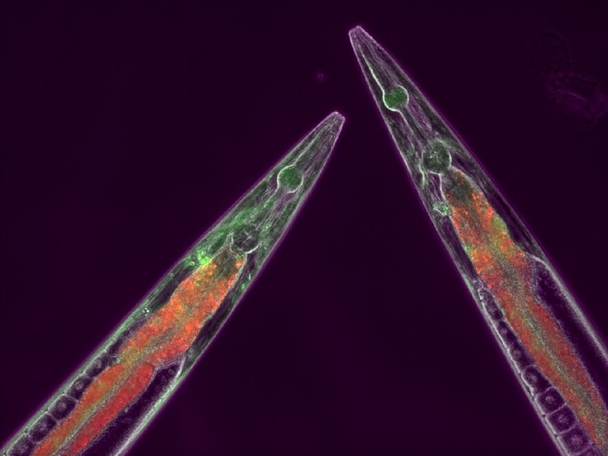
Nematodes, fluorescent microscopy. Photo: Lisa Rossbach
Mouse kidney, confocal laser scanning micrograph
Mouse kidney, confocal laser scanning microscope. Photo: Imaging Centre

Follow us on Instagram for more images.